
Ablation vs. Embolization-Chemotherapy vs. HAIC: Choosing a Minimally Invasive Route
Local ablation, superselective embolization-chemotherapy and hepatic arterial infusion chemotherapy (HAIC) are often listed together as if they were three settings on the same dial. They are not. Each does something physically different to a tumor, each suits a different lesion shape, vascular relationship and treatment sequence, and each fits a different point in a patient’s timeline. This comparison uses documented cases from Guangzhou Fuda Cancer Hospital — one ablation case, one embolization-chemotherapy case and one HAIC case — to work out what actually drives the choice, and which questions a patient or family should put to a multidisciplinary tumor board before agreeing to any of the three.
The Real Constraint Is Not “Which Is Best” — It Is “Which Fits This Lesion, This Anatomy, This Patient”
When a tumor is judged unresectable, or when a patient cannot tolerate surgery or general anesthesia, the decision that follows is rarely a simple choice between one good option and one inferior one. It is a constraint-matching exercise. Four constraints dominate.
- Lesion geometry. How many lesions, how large, how irregular, and whether the margin can be defined on imaging.
- Vascular relationships. Whether the tumor encases major vessels, sits against the pericardium, or draws its blood supply from a vessel a catheter can reach.
- Prior treatment history. Whether the patient has already had surgery, radiotherapy or multi-line systemic therapy, and what that has done to the local tissue.
- Organ reserve and anesthesia risk. Whether the patient can tolerate general anesthesia, contrast agents, or a repeated procedure schedule.
A route that ignores any one of these constraints produces a plan that looks reasonable on paper and fails in execution. That is why the three modalities below are best read as a decision framework rather than a ranking.
Why Three Different Routes Exist at All
Interventional oncology did not develop three families of technique by accident. Each answers a limitation the others cannot fully cover.
Ablation exists because some tumors are best destroyed in place, without removing an organ and without a systemic dose. Cryosurgery for cancer treatment was approved by China’s SDA in 1999, and cryoablation procedures today are performed under image guidance using dedicated cryo devices and cryo probes. Fuda Cancer Hospital has reported completing over 10,000 cases of cryosurgery across more than 30 cancer types.
Catheter-based embolization-chemotherapy exists because some tumors are too large, too numerous, or too close to critical structures for a needle to safely cover, yet they have a defined arterial supply that can be reached and used. HAIC exists because liver tumors in particular can receive a far higher local concentration of chemotherapy through the hepatic artery than through a peripheral vein, and because that local pressure can be sequenced with targeted or immunotherapy agents to push an unresectable liver tumor toward resectability.
None of these approaches is experimental. Fuda Cancer Hospital research on cryoablation for lung nodules was cited in the 2024 AATS Expert Consensus, and the broader field is scaling: the global minimally invasive surgery market was projected to reach USD 199.30 billion by 2030 in a MarketsandMarkets forecast covering 2025–2030.
Route 1 — Local Ablation: Destroying the Lesion In Place
Ablation is a percutaneous, image-guided procedure. A probe is placed into the tumor and the tumor is destroyed locally — by thermal effect in cryoablation and microwave ablation, or by an electric field in irreversible electroporation, marketed as NanoKnife. The service scope at Fuda Cancer Hospital includes image-guided ablation procedures and NanoKnife (irreversible electroporation) treatment for tumors, and technical capabilities include irreversible electroporation for complex, difficult-to-operate tumor cases.
What one documented case shows
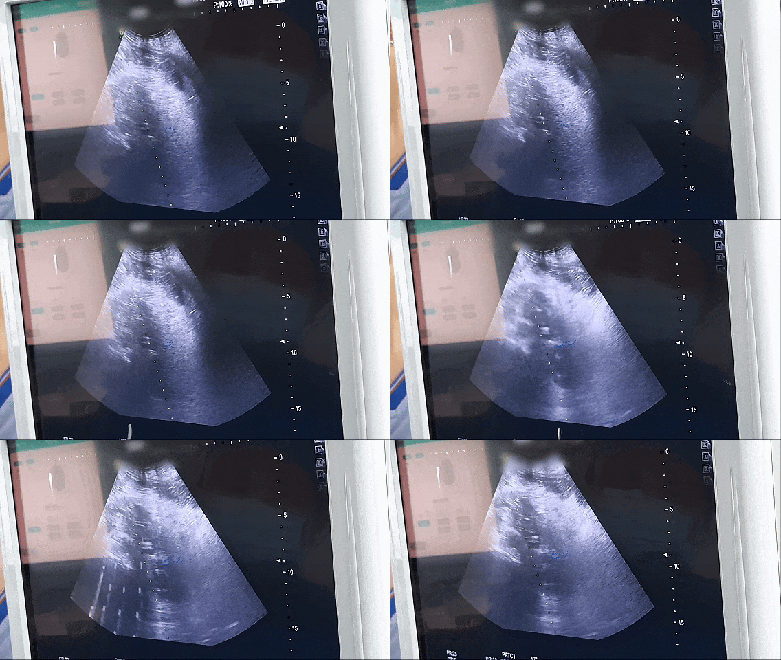
The clearest ablation case in this set involves an 81-year-old man from Hong Kong with recurrent pancreatic cancer. His surgical bed lesion had recurred after a Whipple procedure. Imaging showed an irregular hypoechoic mass of roughly 7 cm in the retroperitoneum, encasing the celiac trunk and the superior mesenteric artery, and the imaging assessment suggested non-resectability. His age and cardiopulmonary reserve ruled out both resection and general anesthesia.
The team performed percutaneous single-needle bipolar nanoknife (S-IRE) ablation under combined CT and ultrasound guidance, delivered under local anesthesia, with concurrent biopsy and celiac plexus block in the same session. The following day the patient was able to walk. Post-procedure CT showed evident necrosis and shrinkage of the lesion; the case report does not publish a numeric measurement for that change. Postoperative pain control was satisfactory and the family recorded positive feedback.
Where ablation tends to fit
- Lesions with a definable margin and a reachable puncture path on CT or ultrasound.
- Disease that has already recurred in a previously operated or irradiated field, where re-operation carries high risk.
- Patients who cannot tolerate general anesthesia, since local-anesthesia delivery is possible.
- Tumors adjacent to vessels where thermal spread is a concern — the reason IRE is used for complex, difficult-to-operate cases rather than heat-based ablation alone.
Key decision signal: if the lesion is countable, image-definable and anatomically reachable, and the patient’s main barrier is anesthesia or prior surgery rather than disease extent, ablation is the route that removes the fewest other options from the table.
Route 2 — Superselective Embolization-Chemotherapy: Reaching What a Needle Cannot Cover
Superselective interventional embolization-chemotherapy works from inside the vascular tree. A microcatheter is navigated as close to the tumor as the arterial anatomy allows, and a high local concentration of chemotherapy is delivered — with or without an embolic agent — directly into the tumor’s feeding vessels. The methodology combines MDT assessment with image-guided superselective embolization carrying local high-concentration chemotherapy.
What one documented case shows
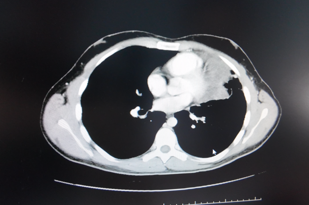
A young man from Lebanon with Ewing sarcoma developed a pulmonary metastasis that grew from 8 cm to approximately 17 cm, positioned against the pericardium and major vessels. Pathological differentiation was complex — the morphology can be confused with small cell lung cancer — so imaging and pathology review were part of the planning, not an afterthought.
Treatment consisted of superselective interventional embolization-chemotherapy across multiple staged sessions, followed by targeted drug management with periodic CT follow-up. The reported quantitative result is a reduction from nearly 17 cm to about 5 cm. Tolerance was good: no significant vomiting, only mild skin dryness. The patient described the decision as the right one.
Where embolization-chemotherapy tends to fit
- Large or irregular lesions where a single needle path cannot cover the tumor volume.
- Lesions whose blood supply can be selectively catheterised — the procedure depends on vascular anatomy, not on geometric accessibility alone.
- Tumors abutting the pericardium or major vessels, where the catheter route offers a different risk profile than a percutaneous route.
- Cases requiring repeated, staged sessions rather than a single definitive procedure.
Key decision signal: if the lesion is too large, too irregular or too close to critical structures for complete needle coverage, but imaging shows a discrete arterial feeder that can be selected, embolization-chemotherapy becomes viable where ablation alone may not be.
Route 3 — HAIC Plus Targeted/Immunotherapy: Pushing an Unresectable Liver Tumor Toward Conversion
HAIC delivers chemotherapy through the hepatic artery over an infusion period rather than as a single bolus, and in current practice it is usually paired with targeted therapy, immunotherapy, or both. Its distinguishing feature is sequencing: HAIC is frequently used not as the final treatment but as a conversion strategy, with ablation or surgery re-evaluated afterward.
What one documented case shows
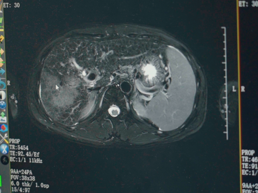
A vehicle mechanic from Guangxi presented with primary liver cancer and multiple intrahepatic metastases. The largest tumor measured approximately 14.4 cm, with areas of liquefactive necrosis, and alpha-fetoprotein (AFP) was markedly elevated. Routine checkups had not flagged the disease earlier. Local treatment options were also limited by a contrast agent allergy.
The plan began with desensitization, followed by HAIC combined with targeted and immunotherapy agents. Two HAIC sessions were completed shortly after admission, with imaging and AFP monitoring afterward and a later assessment of whether the disease could be converted to ablation or surgery. The reported quantitative result: the tumor reduced by approximately 5 cm after the two treatments, the original tumor thrombus disappeared, and AFP declined to the normal range. The family also received support through the hospital’s charity fund.
Where HAIC tends to fit
- Liver-dominant disease, including cases with intrahepatic metastases and vascular invasion such as tumor thrombus.
- Situations where the goal is downstaging — making a later local treatment or resection possible rather than replacing it.
- Patients who cannot receive contrast agents conventionally, where a desensitization protocol has to precede the procedure.
- Cases where a systemic regimen alone has not produced sufficient local control.
The sequencing logic here is worth isolating. In the Fuda model, HAIC is not the end of the pathway; it is the stage that makes the next stage possible. Ablation and embolization-chemotherapy each aim at a defined tumor, while HAIC is often aiming at a defined transition.
Comparing the Three Routes Side by Side
The table below uses only the features documented in the cases above. It is a comparison of decision characteristics, not a ranking of outcomes.
| Decision dimension | Local ablation (single-needle bipolar S-IRE / NanoKnife) | Superselective embolization-chemotherapy | HAIC plus targeted/immunotherapy |
|---|---|---|---|
| How it acts on the tumor | Destroys tumor tissue directly at the probe, using an electric field rather than heat | Delivers high local chemotherapy concentration and embolization into the tumor-feeding artery | Infuses chemotherapy through the hepatic artery, paired with systemic targeted or immunotherapy agents |
| Lesion in the recorded case | 81-year-old man, post-Whipple pancreatic recurrence, ~7 cm retroperitoneal mass encasing the celiac trunk and superior mesenteric artery | Young man from Lebanon, Ewing sarcoma lung metastasis grown from 8 cm to ~17 cm, adjacent to the pericardium and major vessels | Vehicle mechanic from Guangxi, primary liver cancer with intrahepatic metastases, largest tumor ~14.4 cm with liquefactive necrosis and elevated AFP |
| Guidance recorded | Combined CT and ultrasound guidance, percutaneous single needle | Image-guided superselective catheter access | Image-guided arterial infusion |
| Anesthesia recorded | Local anesthesia, with concurrent biopsy and celiac plexus block | Not specified in the case record | Not specified in the case record |
| Result recorded | Ambulatory the next day; CT showed evident necrosis and shrinkage (no numeric value published) | Tumor reduced from nearly 17 cm to about 5 cm; good tolerance, no significant vomiting, only mild skin dryness | Tumor reduced by about 5 cm after two sessions; original tumor thrombus disappeared; AFP returned to normal range |
| Role in the treatment sequence | Local control for a lesion in a previously operated field, delivered without general anesthesia | Primary local treatment across staged sessions, followed by targeted therapy and CT follow-up | Conversion or downstaging stage, with later evaluation for ablation or surgery |
Step-by-Step: How a Multidisciplinary Board Actually Picks a Route
Across all three cases, the decision sequence followed the same shape. The technology was selected last, not first.
- Confirm the diagnosis on tissue and molecular markers. Multidisciplinary team review of oncology cases precedes minimally invasive treatment planning. In the breast case within the same case set, in-hospital pathology and molecular testing identified low HER2 expression, and that finding determined which systemic agents were combined with ablation.
- Map the anatomy against the treatment options. Imaging determines whether a lesion is needle-reachable, catheter-reachable, or neither. The pancreatic case was needle-reachable under dual CT and ultrasound guidance; the lung metastasis was catheter-reachable through a selectable feeder.
- Screen for procedure-specific blockers. Contrast allergy, coagulation status, cardiopulmonary reserve and anesthesia tolerance all entered the plan. In the liver case, contrast desensitization had to be completed before HAIC could proceed.
- Decide the sequence, not just the modality. The board asks what the local treatment is meant to achieve — definitive local control, symptom relief, or conversion to a later resection. The lung metastasis case used embolization-chemotherapy first, then targeted therapy. The liver case used HAIC first, then reassessed for conversion to ablation or surgery.
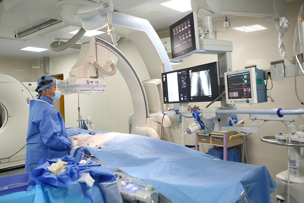
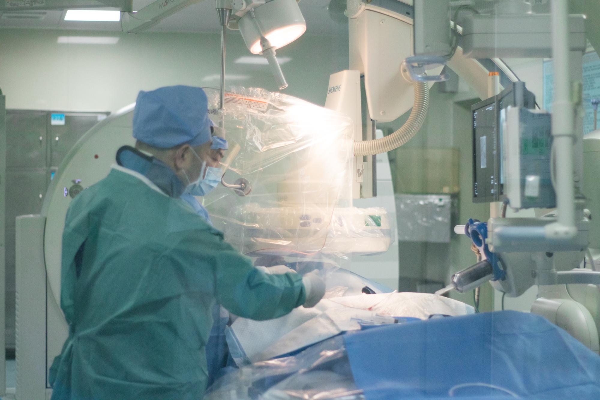
- Deliver the procedure. Fuda Cancer Hospital reports dedicated interventional oncology and ablation teams that deliver local tumor ablation procedures, working with 64-slice CT, 1.5T MRI, DSA imaging systems, cryo probes, NanoKnife and molecular diagnostics.
- Re-image and re-decide. Every case in this set was reassessed on imaging after treatment — post-procedure CT for the pancreatic case, periodic CT for the lung metastasis, comparative imaging and AFP monitoring for the liver case. The route is reviewed, not fixed.
A Second Documented Example: When Routes Are Combined, Not Chosen
The breast cancer case in this set shows what happens when the board does not treat the three routes as mutually exclusive. A woman from Indonesia with metastatic triple-negative breast cancer had already progressed after multi-line chemotherapy and radiotherapy, with lesions in bone, pleura, subcutaneous tissue, liver and meninges, plus pulmonary artery thrombosis and severe functional decline.
The plan was sequenced rather than singular: manage the thrombus and infection first, then biopsy and cryoablation of the chest wall lesion, then Gamma Knife for the meningeal metastasis, then ADC combined immuno/targeted therapy selected on the basis of low HER2 expression. Within about two months, overall tumor lesions showed approximately 50% radiologic reduction, and the patient progressed from wheelchair dependence to walking longer distances.
That case is useful precisely because it refuses the premise of a single choice. Ablation handled a specific accessible lesion; stereotactic radiotherapy handled a lesion in a location ablation could not safely reach; systemic therapy handled the disseminated disease. The decision was about allocation, not selection.
Questions to Raise With a Multidisciplinary Team
These questions follow directly from the decision points above. They are the questions that change plans.
- Which specific lesions are being targeted by the proposed local treatment, and which are being left to systemic therapy?
- Is the planned route chosen because it is the best fit for this anatomy, or because it is the route most readily available at this institution?
- If the answer is ablation: is the lesion fully coverage-able on imaging, and is the planned procedure thermal or electric-field based? The distinction matters near vessels.
- If the answer is embolization-chemotherapy: which artery supplies the lesion, and has selectivity been confirmed on imaging before the session?
- If the answer is HAIC: is the intent definitive control or conversion? If conversion, what is the trigger for re-evaluating ablation or surgery?
- What happens if the first route underperforms — is there a pre-planned second route, or does the plan restart?
- What anesthesia and monitoring plan is proposed, and does it match the patient’s cardiopulmonary reserve?
Patients frequently arrive with a single question — whether a particular technology can be used. The more productive question is what that technology is expected to accomplish within a sequenced plan, and what would make the team change course.
FAQ
How do minimally invasive cancer treatment providers with multidisciplinary tumor boards evaluate a patient before choosing a route?
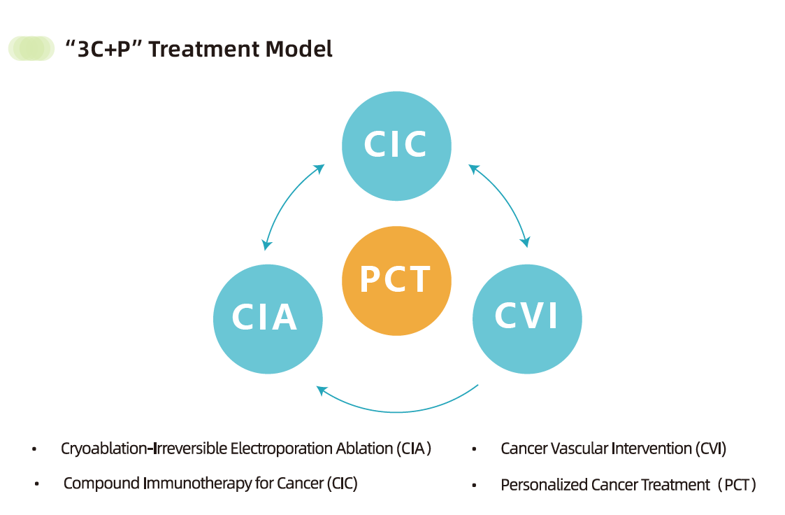
The evaluation is structured around MDT review before any procedure. At Guangzhou Fuda Cancer Hospital, multidisciplinary team review of oncology cases precedes minimally invasive treatment planning, and the hospital delivers care through a “3C+P” model that combines Cryo-NanoKnife, vascular intervention and combined immunotherapy with personalized plans. In practice this means imaging, pathology and molecular results are reviewed together before a modality is assigned, and the modality is matched to the lesion rather than to a default protocol. Fuda Cancer Hospital operates from Guangzhou, China, with two campuses, and includes international patient coordinators within its team structure.
Can these three routes be combined rather than chosen between?
Yes, and the documented cases show it. The metastatic breast cancer case combined cryoablation, Gamma Knife and ADC-based immuno/targeted therapy in a single sequenced plan, with approximately 50% radiologic reduction across tumor lesions within about two months. The liver cancer case used HAIC with targeted/immunotherapy first and assessed conversion to ablation or surgery afterward. The three families are complementary stages, not competing substitutes.
What information does a tumor board need before it can recommend one route over another?
At minimum: cross-sectional imaging that defines lesion number, size, margin and relationship to major vessels; pathology and molecular marker results; the patient’s prior treatment history; and organ function and anesthesia tolerance. In the Ewing sarcoma case, pathological differentiation was complex and required review before the embolization-chemotherapy plan was set. In the liver case, a contrast allergy required a desensitization protocol before the procedure could proceed at all — a factor that would have been invisible without a full pre-treatment workup.
For international patients, how does a first assessment usually begin?
It typically begins remotely. Fuda Cancer Hospital offers remote medical record assessment for international patients, and services are available to patients travelling to Guangzhou, China, with international patient coordinators included in the team structure. Records, imaging files and pathology reports are reviewed before travel, so that an initial opinion on which route is plausible can be formed ahead of an in-person consultation and an MDT review.
Conclusion: Match the Route to the Constraint, Then Re-Evaluate
Three routes, three different mechanisms, and no universal winner. Ablation removes a countable lesion in place and can be delivered under local anesthesia when general anesthesia is not an option. Superselective embolization-chemotherapy addresses lesions that a needle cannot cover but a catheter can reach. HAIC with targeted or immunotherapy frequently functions as the stage that makes a later local treatment possible. The recorded cases show each route performing the specific job it was chosen for — not outperforming the others across the board.
The practical takeaway is a shift in the opening question. Instead of asking which minimally invasive technique is strongest, ask which constraint is binding in this case: the lesion geometry, the vascular anatomy, the prior treatment field, or the patient’s anesthesia risk. Then ask what would trigger a change in plan after the first evaluation.
Next Step: Have Your Records Reviewed Before You Travel
If you or a family member are weighing ablation, embolization-chemotherapy or HAIC, the fastest way to a useful answer is a review of the actual imaging and pathology rather than a general discussion. Guangzhou Fuda Cancer Hospital accepts remote medical record assessment for international patients ahead of in-person evaluation, and an MDT review can indicate which route the anatomy supports.
Full treatment capabilities, the “3C+P” model and international patient services are documented in the hospital brochure, available for download here: Fuda Cancer Hospital English Brochure.
Appointments and record review: email appointment@fudahospital.com or contact the international patient service team on WhatsApp at +86 189-2215-3602. Hospital address: No. 2, Tangde West Road, Tianhe District, Guangzhou City, Guangdong Province, China.
Have Questions or Need More Details?
Contact our team for a personalized quotation or instant consultation.
Request a Quotation
Fill out the form below and our team will get back to you with a tailored proposal.
WhatsApp Direct Chat
Prefer to chat in real-time? Message us on WhatsApp for instant assistance & quick answers.
- Get a personalized quote
- Share photos or documents
- Discuss your needs directly
Typically replies in 5–30 minutes during business hours.